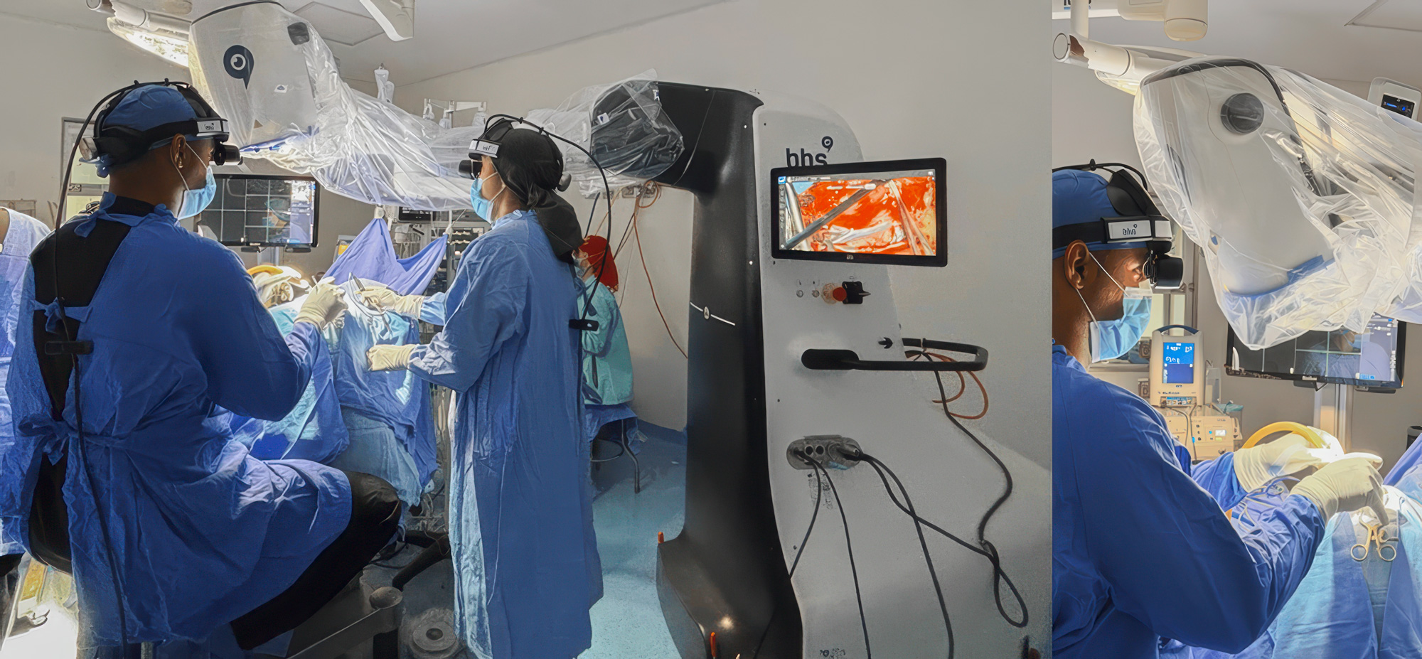
A recently published paper presents a minimally invasive approach to frontal convexity meningioma resection using the RoboticScope.

New Case Report on Robotic Meningioma Surgery
Minimally invasive techniques continue to shape modern neurosurgery. By reducing surgical exposure while maintaining precision and control, these approaches aim to minimize tissue trauma, support faster recovery, and improve the overall surgical workflow. As procedures become more refined, advanced visualization technologies are playing an increasingly important role in helping surgeons work safely within smaller surgical corridors.


One example is highlighted in a recently published case report in Cureus by Marco A. Baralt Ramirez, Adriel Lopez Salazar, Floricel Olimpia Villegas Amador, Gianfranco Stefanoni and Ashanti Gutiérrez Quintanar. The publication describes how the RoboticScope was used to support a minimally invasive approach for the resection of a frontal convexity meningioma, demonstrating the role of robotic visualization throughout the procedure.

A minimally invasive procedure in practice

The case involved a 23 year old patient who was diagnosed with a left frontal convexity meningioma following imaging after a sports related head injury. Due to the tumor's superficial location and well defined anatomical boundaries, the surgical team selected a minimally invasive approach combined with neuronavigation and robotic visualization.


The procedure was performed through a 4 cm skin incision and a 3 cm craniotomy. Using the RoboticScope, the surgeons carried out microsurgical dissection while preserving the surrounding cortical anatomy and venous structures. Complete Simpson Grade I tumor resection was achieved, with histopathology confirming a WHO Grade I fibrous meningioma.

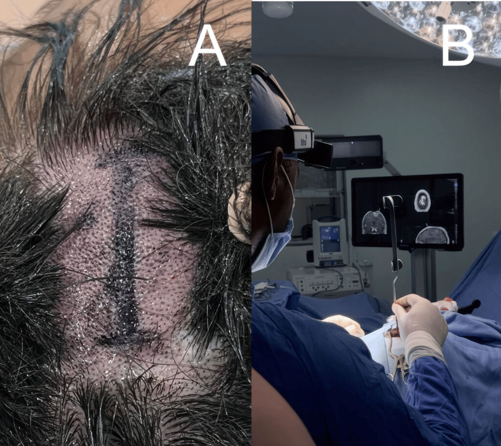
Minimally invasive frontal approach and neuronavigation planning (A) Preoperative marking of a 4 cm minimally invasive frontal incision with preservation of the patient's hair. (B) Intraoperative neuronavigation confirming the planned minimally invasive frontal entry point and surgical trajectory. Baralt Ramirez M A, Lopez Salazar A, Villegas Amador F, et al. (July 09, 2026) Robotic Minimally Invasive Surgery for Frontal Convexity Meningioma: A Case Report and Technical Note. Cureus 18(7): e112373. doi:10.7759/cureus.112373

Robotic visualization throughout the procedure

Throughout the procedure, the RoboticScope supported continuous visualization of the surgical field. Its intuitive Head Gesture Control allowed the lead surgeon to adjust zoom, focus, illumination, and viewing angle without interrupting the surgical workflow. This supported precise microsurgical dissection within the limited surgical corridor while helping maintain an ergonomic working position and continuous focus on the procedure.

Baralt Ramirez M A, Lopez Salazar A, Villegas Amador F, et al. (July 09, 2026) Robotic Minimally Invasive Surgery for Frontal Convexity Meningioma: A Case Report and Technical Note. Cureus 18(7): e112373. doi:10.7759/cureus.112373

What this case demonstrates

The surgeons describe robot assisted minimally invasive surgery as a promising option for selected frontal convexity meningiomas. They note that combining robotic visualization, neuronavigation and minimally invasive surgical techniques may help reduce brain manipulation, surgical morbidity, postoperative pain and recovery time compared with conventional transcranial approaches in appropriately selected cases.


This case demonstrates how robotic visualization can support minimally invasive cranial procedures by enabling precise microsurgical workflows in carefully selected patients.

Read the full case report here.


Baralt Ramirez M A, Lopez Salazar A, Villegas Amador F, et al. (July 09, 2026) Robotic Minimally Invasive Surgery for Frontal Convexity Meningioma: A Case Report and Technical Note. Cureus 18(7): e112373. doi:10.7759/cureus.112373